A research team at UHN's Donald K. Johnson Eye Institute in collaboration with the Institute of Biomedical Engineering at the University of Toronto (U of T) has discovered that transplanted retinal cells can share essential materials with host cells in the lab, offering a promising avenue for delivering therapies directly to damaged areas of the eye.
The degeneration of photoreceptors, a leading cause of blindness affecting one in 2000 individuals globally, stems from the loss of light-sensing cells in the eyes. It is an irreversible condition due to the limited regenerative capacity of our central nervous system.
Cell therapy has been explored as a potential vision restoration treatment, involving the introduction of healthy donor cells into the retina to form new connections with surrounding neurons.
However, studies have shown that transplanted cells rarely form new connections. Instead, they transfer cellular materials, such as proteins, into the host retina in a process called material transfer.
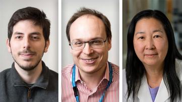
“This bidirectional process has been observed mainly in experimental models,” explains Dr. Margaret Ho, a Vanier scholar, a PhD Candidate at the Institute of Biomedical Engineering at U of T and first author of this study. “However, until now, it has remained unclear whether material transfer can occur between human cells.”
"We want to understand if human photoreceptors derived from stem cells can engage in material transfer, as this could be a promising pathway for targeted drug delivery to diseased cells," explains Dr. Valerie Wallace, Senior Scientist at Krembil Research Institute and a senior author of this study.
This collaborative project draws on the complementary expertise of Dr. Valerie Wallace and Dr. Molly Shoichet. Dr. Wallace specializes in material transfer, experimental models, and tissue analysis, while Dr. Shoichet focuses on in human stem cell growth and manipulation, and the creation of photoreceptors from retina tissue-like structures called retinal organoids.
"This work couldn't have been possible without the collaboration between our labs,” said Dr. Molly Shoichet, a professor at the University of Toronto and one of the corresponding authors of the research.
The researchers used two approaches to understand if human stem-cell-derived photoreceptors can transfer materials to host cells. First, they tested this by transplanting their human donor photoreceptors into experimental models with intact host photoreceptors, and then they tested species specific transfer by co- culturing donor and host cells from dissociated human retinal organoids in a lab dish.
In the first set of experiments, they tried transplanting human photoreceptors from different stages of development into experimental models with eye problems. No transfer was observed, even though both cell types survived in the eye the same amount of time.
In the second experiment in the lab, researchers observed that human donor cells could transfer their content to host cells, which has never been shown before. This phenomenon opens up an entirely new way of thinking about vision repair.
"This result suggests the importance of considering species when designing models to study material transfer in the future," emphasizes Dr. Ho.
"Photoreceptors derived from human stem cells could still hold promise for treating eye diseases in humans. We need to keep studying how to optimize cell transplants to advance treatments for eye disease and improve transplant outcomes," concludes Dr. Wallace.
These findings are a step forward in our understanding of transplant therapy for eye disease. This advancement prompts new questions about whether human eye cells possess the same capabilities, an area largely unexplored due to prior studies focusing on late-stage eye disease models devoid of host cells.
This work was supported by Medicine by Design, The Stem Cell Network, the Natural Sciences and Engineering Research Council, the Canadian Institutes of Health Research, the Vision Science Research Program and UHN Foundation.
Ho, M.T., Kawai, K., Abdo, D. et al. Transplanted human photoreceptors transfer cytoplasmic material but not to the recipient mouse retina. Stem Cell Res Ther 15, 79 (2024). https://doi.org/10.1186/s13287-024-03679-3
This year, at the 50th annual Gallie Day, Dr. Anna Gagliardi, Senior Scientist at UHN’s Toronto General Hospital Research Institute, was honoured with the prestigious Lister Prize.
Gallie Day is an annual event hosted by the Department of Surgery at the University of Toronto to honour and recognize outstanding achievements within the surgical community. The Lister Prize, the most prestigious research award in the Department of Surgery, celebrates an investigator who has shown outstanding and continued productivity of international stature as evidenced by research publications, grants, mentorship, and beyond.
Dr. Gagliardi's accolade stands as a testament to her unwavering dedication and impactful contributions to surgical research. Through her innovative efforts, she has not only helped advance the frontier of knowledge but has also profoundly impacted patient care and outcomes. Her work, spanning decades, has been a beacon of inspiration for colleagues, students, and collaborators alike.
Reflecting on the award, Dr. Gagliardi shares, “It is an immense honour to be recognized in this way for the many years of dedicated research—a journey that has been both a profession and a passionate pursuit. This award is a testament to the collective efforts of all those who have supported and inspired me throughout this transformative journey, including esteemed colleagues, diligent students, and invaluable collaborators.”
Dr. Gagliardi’s work focuses on optimizing the implementation of guidelines, organization, and individual patient and family engagement as well as person-centered care for women. At the University of Toronto, she is a Professor in the Department of Surgery, the Institute of Health Policy, Management and Evaluation as well as the Institute of Medical Science.
Researchers from UHN’s Krembil Brain Institute have delved into the intricate relationship between neuron hyperactivity and the accumulation of amyloid-β protein, unveiling promising pathways for early diagnosis and intervention of Alzheimer’s disease.
Alzheimer’s is a slow-progressing disease that can start years before any noticeable symptoms appear. Without any known cure, early detection and treatment of the disease is critical. One key player identified in the progression of Alzheimer’s is amyloid-β (Aβ), a protein that accumulates in the brain and triggers excessive neuronal activity, which has been associated with the development of the disease.
A team led by Dr. Maurizio De Pittà, Krembil Brain Institute Scientist and senior author of the study, applied mathematical models to explore the relationship between Aβ and excessive neuronal activity.
“In this study, we used mathematical models to mimic the intricate processes underlying Alzheimer’s,” explains Dr. De Pittà. “These models enable us to simulate, analyze, and predict the interaction between Aβ and neuronal activity over time.”
Researchers found a complex relationship between Aβ and neuronal activity. A build-up of Aβ initiates a cascade of events that leads to excessive neuronal activity. This heightened activity, in turn, can also increase Aβ production, establishing a cycle that drives disease progression.
The researchers highlighted that the relationship between Aβ accumulation and neuronal hyperactivity differs from one brain region to another. Thus, understanding variations in brain activity across different regions can provide valuable insights into the diverse and intricate manifestations of Alzheimer’s disease.
“By unravelling the complex interactions in the brain, we can identify new avenues for early detection and targeted treatment,” concludes Dr. Giulio Bonifazi, a previous PhD student in Dr. De Pittà’s lab and first author of the study. “With further research and clinical trials, these findings hold promise for transforming the diagnosis and management of Alzheimer’s disease.”
This work was supported by “la Caixa” foundation, the Krembil Foundation, BIOEF – Basque Foundation for Health Innovation and Research, the Centro de Investigación Biomédica en Red Enfermedades Neurodegenerativas (CIBERNED) and UHN Foundation. Dr. Maurizio De Pittà is an Assistant Professor in the Department of Physiology at the University of Toronto.
Bonifazi G, Luchena C, Gaminde-Blasco A, Ortiz-Sanz C, Capetillo-Zarate E, Matute C, Alberdi E, De Pittà M. A nonlinear meccano for Alzheimer's emergence by amyloid β-mediated glutamatergic hyperactivity. Neurobiol Dis. 2024 May. doi: 10.1016/j.nbd.2024.106473.
A new study from UHN’s Toronto General Hospital Research Institute (TGHRI) has identified the impact of frailty on hospitalized patients with interstitial lung diseases (ILD).
Interstitial lung diseases are a group of lung disorders that cause progressive scarring and declining lung function. Every year, 3% to 14% of patients with ILD suffer acute exacerbations—flare-ups (AE-ILD), which are associated with high morbidity and mortality. Identifying risk factors is crucial for predicting outcomes, allocating resources, assessing transplant eligibility, and optimizing recovery.
“Frailty is a syndrome characterized by the reduced reserve of energy to maintain functions and cope with stressors that happens as we age,” says Dr. Dmitry Rozenberg, Scientist at TGHRI and senior author of the study. “Frailty is often associated with greater risks in chronic lung diseases and has also been associated with diminished quality of life and transplant-free survival in patients with chronic respiratory diseases.”
Although it is known that frailty can be an indicator of outcome for patients with ILD and that approximately 50% of ILD patients experience frailty, the implications of frailty during acute exacerbations of ILD have remained largely unexplored.
“Our study aimed to understand the association between frailty and various aspects of AE-ILD including clinical characteristics, physical function, hospital outcomes, and post-AE-ILD recovery,” says Dr. Marine Van Hollebeke, former Postdoctoral Researcher at UHN and co-first author of the study. Dr. Van Hollebeke was co-supervised by Dr. Rozenberg and Dr. W Darlene Reid, Senior Scientist at KITE, Professor at the University of Toronto (U of T), and co-author on the study.
The team analyzed retrospective data spanning from January 2015 to October 2018. They examined 89 patients hospitalized with AE-ILD, 31 of them being identified as having frailty based on a previously developed index. This indicates that frailty is prevalent, being present in one-third of patients admitted with AE-ILD.
“We found that patients with frailty tended to be older, had a higher burden of comorbidities—for example, diabetes or cardiovascular disease, exhibited reduced physical function prior to hospitalization, and decreased independence,” says Dr. Karan Chohan, former medical student at U of T and co-first author of the study. “Patients with frailty also had more major medical complications (32% of patients with frailty vs 10% without) and required more multidisciplinary support during hospitalization.”
However, the study also showed that frailty was not associated with one-year mortality when factoring in lung transplantation, which is a life-saving procedure.
This study underscores the importance of identifying frailty in ILD patients, as it may help health care providers better understand prognosis, allocate resources more effectively, and tailor care plans to address the specific needs of individuals.
Future research will evaluate if frailty can be modified prior to or during hospitalization with AE-ILD and whether offering rehabilitation strategies during or after hospitalization can improve hospital and post-discharge outcomes.
This work was supported by the Canadian Pulmonary Fibrosis Foundation, the Canadian Institutes of Health Research (CIHR), the Temerty Faculty of Medicine, U of T, and UHN Foundation.
Dr. Dmitry Rozenberg is an Assistant Professor in the Temerty Faculty of Medicine at U of T.
For competing interests of co-authors, please see manuscript.
Van Hollebeke M*, Chohan K*, Adams CJ, Fisher JH, Shapera S, Fidler L, Goligher EC, Martinu T, Wickerson L, Mathur S, Singer LG, Reid WD, Rozenberg D. Clinical implications of frailty assessed in hospitalized patients with acute-exacerbation of interstitial lung disease. Chron Respir Dis. 2024 Jan-Dec;21:14799731241240786. doi: 10.1177/14799731241240786.
*Both authors contributed equally to this work
Frailty is associated with greater risks in chronic lung diseases, including the need for hospitalization and higher mortality rates.
Researchers at UHN’s KITE Research Institute have recently leveraged the power of deep learning to improve nerve signalling interpretation, paving the way for more effective neuroprosthetic devices and offering new hope for those with impaired motor function.
Neurological injuries and amputation frequently result in reduced motor function and diminished quality of life. Neuroprosthetics are small, implanted devices designed to help restore lost movement or sensory function by using electrical impulses to stimulate nerves—a process known as neurostimulation.
These devices use small electrodes to capture sensory information from the nerves and the environment to refine movements. However, interpreting these nerve signals is challenging due to the nature of the electrodes. Placing electrodes inside the nerve provides clearer signals but poses a risk of neural tissue damage. Electrodes positioned outside the nerves are less invasive but yield lower signal quality.
Dr. José Zariffa, Senior Scientist at the KITE Research Institute and senior author of the study, led a team that applied deep-learning models to enhance nerve signal interpretation.
“Deep learning is a form of artificial intelligence that excels at analyzing complex data and recognizing patterns,” explains Dr. Zariffa. “By applying these models, we can decipher complex and low-quality nerve signals and extract more precise information.’
To evaluate the effectiveness of these models, the research team tested different deep-learning models on nerve signals collected from the sciatic nerve—one of the largest nerves in the body running from the lower back to the foot. They analyzed the model’s ability to understand the nerve signals and classify them based on where they originated in the body.
“We found that deep learning significantly improved nerve signal interpretation accuracy,” states Aseem Partap Singh Gill, previous undergraduate student in Dr. Zariffa’s lab and first author of the study. “The best-performing model achieved an accuracy of around 93%, demonstrating how deep learning can enhance nerve signalling interpretation and improve neuroprosthetics for individuals living with disabilities.”
Future research aims to apply these models to analyze signals from multiple electrodes, different nerves, and more complex movements.
This work was supported by the Natural Sciences and Engineering Research Council of Canada and UHN Foundation. Dr. José Zariffa is an Associate Professor in the Institute of Biomedical Engineering and is cross-appointed at the Edward S. Rogers Sr. Department of Electrical & Computer Engineering and the Rehabilitation Sciences Institute at the University of Toronto.
Gill APS, Zariffa J. Time series classification of multi-channel nerve cuff recordings using deep learning. PLoS One. 2024 Mar 12. doi: 10.1371/journal.pone.0299271.
Researchers at UHN's Princess Margaret Cancer Centre have discovered a promising approach to boost the effectiveness of immunotherapy cancer treatments by manipulating the metabolism of a specific type of immune cell.
Cytotoxic T cells are a type of white blood cells that play a key role in the surveillance and elimination of abnormal cells, including cancerous ones.
Some tumours can evade T cell recognition and attack, which can lead to disease progression. One common strategy in cancer immunotherapy is Adoptive Cell Therapy (ACT), which involves using a patient's own immune cells to fight cancer.
In ACT, immune cells such as T cells are collected from the patient's blood or tumour tissue, modified or activated to enhance their ability to fight tumours, and then infused back into the patient.
"Although successful in some cases, cell therapy typically doesn't offer long-lasting benefits for most patients," explains Dr. Pamela Ohashi, Senior Scientist at Princess Margaret Cancer Centre and senior author of this study. "We aim to improve the way ACT is made to increase its effectiveness long-term."
Previous research indicates that directing interventions towards metabolic stress pathways, which have been conserved through evolution, like nutrient deprivation or energy production, can enhance the ability of T cells to control tumours.
The team set out to study an important molecule that cells produced in response to nutrient starvation, called GCN2 (kinase general control non-depressible 2).
"We knew that when GCN2 is activated, it generally reduces overall protein production, leading to the expression of molecules that coordinate various cellular activities, like protein uptake," explains Dr. Michael St. Paul, Post-Doctoral researcher at Princess Margaret Hospital and first author of this study. "However, the specific role of GCN2 in cytotoxic T cells remains unclear."
To study this, researchers started by investigating amino acid depletion, specifically the lack of arginine, which is recognized for its crucial role in immune cell function.
By moving activated cytotoxic T cells from standard medium to one depleted of arginine, scientists noted an increased activation of GCN2-induced stress response. This was shown by an increase in the production of immune-regulating molecules (cytokines) and enhanced energy production through oxidative metabolism.
To validate these results, they used halofuginone (halo), a GCN2 activator, and observed comparable impacts on T cell function and oxidative metabolism.
"Interestingly, this metabolic shift persisted even after removing the halo from the cells, indicating a sustained change in the metabolism of T cells," explains Dr. Sam Saibil, Staff Oncologist at the Princess Margaret Cancer Centre. "This means that harnessing the intrinsic capabilities of the immune system could lead to more targeted and durable therapeutic interventions."
This study helped understand how GCN2 signaling affects T cell responses to arginine levels, shedding light on potential therapeutic strategies for enhancing immune responses against cancer.
"Our research opens doors to enhancing various forms of cancer immunotherapy by targeting GCN2, signaling a potential paradigm shift in cancer treatment strategies," concludes Dr. Ohashi.
This work was supported by the Canadian Institutes for Health Research Foundation and the Princess Margaret Cancer Foundation.
St Paul M, Saibil SD, Kates M, Han S, Lien SC, Laister RC, Hezaveh K, Kloetgen A, Penny S, Guo T, Garcia-Batres C, Smith LK, Chung DC, Elford AR, Sayad A, Pinto D, Mak TW, Hirano N, McGaha T, Ohashi PS. Ex vivo activation of the GCN2 pathway metabolically reprograms T cells, leading to enhanced adoptive cell therapy. Cell Rep Med. 2024 Mar 19;5(3):101465. doi: 10.1016/j.xcrm.2024.101465.
To bridge education gaps and foster cultural sensitivity, a study from UHN’s The Institute for Education Research (TIER) explored the perceptions of nursing students on the experiences of Indigenous peoples within Canada’s healthcare system. Their results revealed strategies for tailoring nursing education to ensure more culturally safe and compassionate care.
Indigenous communities have long faced health inequities marked by a higher burden of illness, inequitable health factors, and premature mortality. Indigenous peoples encounter discrimination every day, including in healthcare settings.
In 2015, the Truth and Reconciliation Commission of Canada developed 94 calls to action to address the impact of residential schools and promote reconciliation in Canada. This involved urging nursing students to understand Indigenous history and be trained to provide culturally safe care.
The Canadian Association of Schools of Nursing developed strategies to integrate Indigenous health content into nursing education; however, the effectiveness of these educational initiatives remains largely unknown.
Dr. Kateryna Metersky, Affiliate Scientist at TIER and a nurse at UHN’s Toronto Western Hospital, led a research team to explore nursing student knowledge and understanding of the experiences of Indigenous peoples within the Canadian health care system and identify how educators can better equip students.
Fifteen nursing students from a university in Ontario, Canada, discussed and shared reflections on a video from the Aboriginal Peoples Television Network, the first national Indigenous broadcaster in the world, entitled “Indigenous Peoples and the Problems in Health Care”.
In their reflections, students showed a deep understanding of the complex factors impacting Indigenous health, including racism, historical injustices, and structural barriers to accessing healthcare, particularly those living in remote communities.
Students also identified strategies to help provide culturally safe care, such as building trust-based relationships, establishing rapport, eliminating unconscious biases, and developing advocacy skills to challenge social inequities within Canada’s complex healthcare system.
“Examining the educational content of nursing programs is important to assessing the value of education provided and tailoring future curriculum to better address knowledge gaps,” concludes Dr. Metersky. “Future work should be conducted to determine the impact of tailored curriculum on preparing students to become culturally compassionate practitioners.”
This work was supported by UHN Foundation.
Metersky K, Chandrasekaran K, Ezekiel S. Undergraduate Nursing Student Reflections on Indigenous Peoples' Experiences With the Canadian Health Care System. Nurs Educ Perspect. 2024 Mar 7. doi: 10.1097/01.NEP.0000000000001255.
Research conducted at UHN's research institutes spans the full spectrum of diseases and disciplines, including cancer, cardiovascular sciences, transplantation, neural and sensory sciences, musculoskeletal health, rehabilitation sciences, and community and population health.
Learn more about our institutes by clicking below: